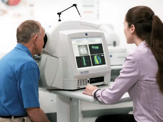
Although the term 'tomography' might evoke the radiation associated with computed tomography in radiology for our patients, OCT operates with light and does not contain any radiation, making it safe for everyone.
The device calculates how strongly and for how long the light waves it sends to the eye are reflected, displaying the tissues inside the eye with better resolution than a microscope. This allows for the visualization of many eye tissues, including the macula and optic nerve, down to the finest detail within a five-second scan, and enables cell counting.
Videos: Prof. Dr. Ahmet AkmanEye Tomography OCTTüm Videolar
Glaucoma, or commonly known as eye pressure disease, is a condition that leads to vision loss due to the death of nerve cells connecting the eye to the brain. For detailed information on this, you can visit our glaucoma page.
OCT imaging shows the status of these connecting cells and indicates whether glaucoma is present or not. Moreover, with repeated scans, it also shows whether the disease is progressing. Along with visual field testing, it is the most important test in the diagnosis and monitoring of glaucoma. Visual field testing only shows damage when 30-40% of nerve cells have died, making it crucial for moderate and advanced stages of glaucoma.
OCT eye tomography can detect damage in the nerve with just 1-2% loss years before visual field deterioration. Therefore, it is crucial for early diagnosis. OCT eye tomography not only enables early diagnosis of eye pressure but also shows that the nerve is normal in many patients, preventing misdiagnosis and unnecessary medication use. Additionally, with repeated scans, it shows the progression of the disease and helps determine if the treatment is sufficient.
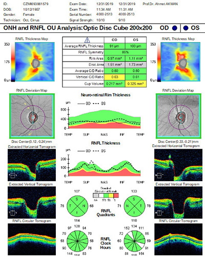 Normal Oct Optic Disc Eye Tomography
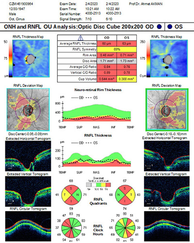 Severe Glaucoma with Oct Eye Tomography
With OCT, it is possible to observe damage to ganglion cells, which are affected cells in glaucoma, at three points. The first and most commonly used method is to measure and evaluate the retinal nerve fiber layer formed by the axons, which are connections of these cells extending to the brain.
The second is to determine the cupping formed by the loss of these axons in the optic nerve. The third and latest method is to directly measure the damage to these cells in the macula.
OCT eye tomography performs all these functions with a 5-second scan and compares the findings with a normal human eye to determine the presence of damage.
An important point here is that due to the unique nature of each person's eye, some normal anatomical differences in some eyes can be misinterpreted as damage by the device. In such cases, avoiding misdiagnosis depends on the doctor's experience with OCT.
Prof. Dr. Ahmet Akman is not only one of the leading experts in our country but also among the world's leading experts. His book "OCT in Glaucoma" is the only reference book on the subject and guides doctors worldwide, especially in the USA and Europe. Additionally, Prof. Dr. Ahmet Akman has been giving a 2-hour course on this topic at the American Academy of Ophthalmology Congress, the world's largest eye disease congress, for the last 4 years and is the only Turkish ophthalmologist to organize his own course at this congress.

Prof. Dr. Ahmet Akman's Glaucoma Oct Book Currently on Sale on Amazon US
After the diagnosis is made or if the diagnosis is in doubt, the initial and final state of the patient are compared with each other with repeated shots on the same device, to determine whether the damage to the ganglion nerve cells is progressing. has it stopped? understandable.
f there is progression, it is understood that the eye pressure needs to be further reduced. If the nerve remains normal and always stays normal, i.e., if there is no cell loss, then the patient is followed without medication, even if the eye pressure is high.
In summary, glaucoma (eye pressure) is a disease that requires monitoring. Especially with OCT devices equipped with a monitoring program, patient follow-up can be done very sensitively. While patients are protected from unnecessary medication use, deteriorating eyes are detected early, making treatments more effective and preventing vision loss.
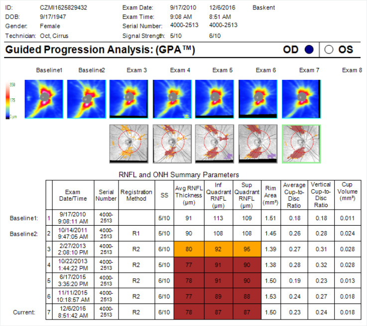
The use of OCT change analysis in follow-up shows the areas where nerve loss occurs in red. This analysis is our most powerful tool in glaucoma follow-up.
In Conclusion: Glaucoma is a disease that requires monitoring. It should be followed by an experienced glaucoma specialist with qualified equipment. Prof. Dr. Ahmet Akman provides services with Zeiss Cirrus OCT, the most advanced device in glaucoma, and Zeiss Humphrey HFA3 visual field device in his clinic. Prof. Dr. Ahmet Akman personally conducts all tests and evaluates them in conjunction with the examination. You can reach the positive comments of many of our patients about glaucoma diagnosis and monitoring from our comments page.

You can call us immediately for detailed information, consultation or appointment.
Contact informationAhmet Akman, MD, FACSProfessor of Ophthalmology